The retina is a sensory membrane that lines the eye and controls how we see our surroundings. A deeper look into this part of the eye can help doctors detect and discover certain health issues.
A good example is the story of Cheree Burnette. Ms. Burnette was experiencing blurred vision and she knew an eye exam could help determine the problem. She told KPLCTV Channel 7 “…I had a problem that caused me to have distorted vision and came in pretty quickly after that and they were able to tell me exactly what I had.”
Ms. Burnette’s optometrist, Dr. Robert Janot, performed a retinal exam using the Daytona ultra-widefield retinal imaging device, which identified Ms. Burnette’s condition as a retinal vein occlusion, or a blocked vein in her eye. The image also helped reveal another problem, which could have been the initial cause of the retinal vein occlusion.
“I had undiagnosed pre-hypertension, which we believe was the cause,” she told KPLCTV Channel 7.
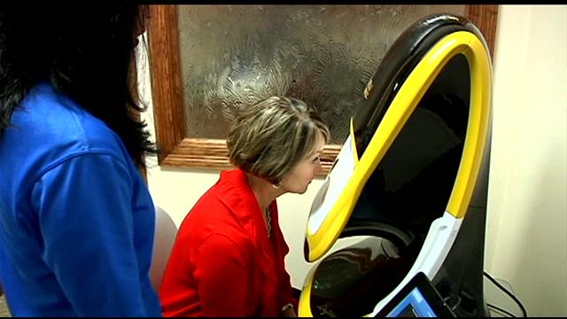
Image from Kplctv.com
Dr. Janot shared that he performs the optomap exam on all patients to check their eye health and see issues that might be occurring beyond the surface. An optomap image provides a panoramic view of the retina, allowing eyecare professionals to look at the only blood vessels in the body that can be seen in a natural state on a deeper level. Dr. Janot says this technology offers a unique look into what’s going on with the retina in terms of diagnosing other health problems. “We’re really privileged to be able to find early signs of systemic diseases through the eye, such as diabetes and high blood pressure,” he said.
And undiagnosed pre-hypertension could very well have been the cause of Ms. Burnette’s retinal vein occlusion – an issue that may have remained unknown for quite sometime without a retinal scan. She now sees close to 20/20 again.
To learn more about the optomap ultra-widefield retinal imaging technology, please visit the Optos website.