Results from a recent publication call for the use of consistent nomenclature when describing the field of view captured by retinal images. The International Widefield Imaging Study Group has proposed the need for consistent nomenclature for widefield and ultra-widefield imaging based on normal anatomic landmarks. When describing the area captured by an imaging modality, it is important to be consistent in meaning so the capabilities of the technology are clear to the reader.
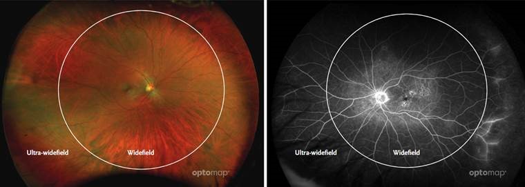
The panel defines ultra-widefield as images showing retinal anatomy anterior to the vortex vein ampullae in all four quadrants. Widefield is defined as an image centered on the fovea and includes the retina in all four quadrants posterior to and including the vortex vein ampullae. The panel recommends this standardized nomenclature for use in future publications1.
Over the last decade, many large studies have underlined the importance of appropriately imaging the periphery to support the detection and management of disease in a variety of areas including telemedicine screening2,3,4, diabetic retinopathy5,6, age-related maculardegeneration7, vascular disease8, pediatric retinal disease9, inflammatory disease10,11,12 and even some systemic diseases. Consistently, optomap imaging has been demonstrated to capture the widest field of view in a single capture of any imaging technology14,15,16,17.
“A single capture image which provides a view of the
vortex veins in all four quadrants and beyond, thus
meeting the widefield & ultra-widefield definitions,
would offer enhanced efficiency in a real-world clinical
setting versus a montage image, whether it be manual or
automated.”
— Netan Choudhry M.D. FRCS(C) DABO
Based on the panel’s findings, a single capture ultra-widefield (UWF™) retinal image, or an image with “no sweeps” can therefore provide enhanced efficiencies for practices and other clinical settings. As the ONLY single-capture UWF image to meet this definition, optomap, is the best choice for increasing efficiencies in your practice or other clinical settings.
Increase your practice efficiency and increase your revenue with optomap. No Sweeps = Increased Efficiency.
Read the full summary here: https://optos.is/NoSweepsUWF and then contact us to find out how to put optomap into your clinical setting.