optomap UWF Advances
A recent review of advances in ultra-widefield retinal imaging systems and applications highlights the importance of visualizing pathology in the retinal periphery specifically related to diabetic retinopathy, retinal vein occlusion uveitis, and the pediatric retina.
UWF Retinal Imaging - An Update on Advances
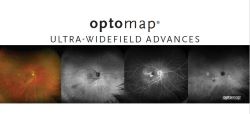
Optos devices capture optomap images, all of which meet the recently defined ultra-widefield (UWF™) definition established by a consensus group of international retinal imaging experts. UWF is a single-capture image that is centered on the fovea and contains anatomical features anterior to the vortex vein ampullae in all four quadrants, where the field of view is 110°- 220°. Optos is currently the only UWF system commercially available on the market that fits this definition. The clinical utility of UWF imaging for diabetic retinopathy, retinal vein occlusion, uveitis, and the pediatric retina continues to improve our understanding, management and treatment of these diseases by capturing peripheral changes in pathology. With the use of multimodal systems, such as those from Optos, an array of capabilities including color imaging, autofluorescence, fluorescein angiography, indocyanine green angiography, and optical coherence tomography are used to identify abnormalities that may have clinically-significant implications.
Read the complete article
Download the Clinical Summary
Contact Optos directly to put advanced UWF retinal imaging devices in your practice or clinical setting.