AMD is a common eye condition that causes damage to the macula, and is a leading cause of vision loss among people age 50 and older. In some people, AMD advances so slowly that vision loss does not occur for a long time. In others, the disease progresses faster and may lead to a loss of vision in one or both eyes. AMD by itself does not lead to complete blindness, with no ability to see. However, the loss of central vision in AMD can interfere with simple everyday activities, such as the ability to see faces, drive, read, write, or do close work, such as cooking or fixing things around the house.
Earlier detection and treatment of AMD can prompt steps to be taken to help reduce vision loss and slow the advance of the disease. Data suggests that the retinal periphery can exhibit some important morphological changes, such as peripheral drusen and reticular pigmentary changes, which are frequently connected with the wet form of AMD. Typically, disease progression has been documented using fundus cameras that image only about 45-50% percent of the retina. By using UWF for AMD evaluation, over 80% of the retina is now analyzed to record peripheral fluorescein angiographic changes in AMD patients.
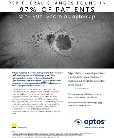
The 12-year follow-up of a study known as the Reykjavik Eye Study evaluated subjects with optomap color and autofluorescence imaging and found that 67% of subjects in the study had peripheral AMD-like changes. Additionally, a subsequent study, knowns as the OPERA (Optos Peripheral RetinA) study found that peripheral retinal changes were more prevalent in eyes with AMD than in those without. Drusen and reticular changes were evident in 97% of the eyes with AMD in both the mid and far periphery. These studies go to show that AMD may be more than a “macular” condition but one that involves the entire retina. Other research also shows that optomap icg captured significant peripheral changes in 80% of AMD patients. These studies all contribute to the benefits of ultra-widefield imaging in the diagnosis and management of AMD. Longitudinal studies of peripheral changes in AMD and their impact on visual function are underway to further understanding of the disease. contact us to learn how ultra-widefield optomap imaging can assist eyecare professionals to find and document AMD earlier and potentially change the course of the disease.