Nearly 30,000 sports-related eye injuries are treated in U.S. emergency rooms each year. The good news is that 90% of serious eye injuries can be prevented by wearing appropriate protective eyewear. Because such a high percentage of sports-related eye injuries are preventable, the organization Prevent Blindness has dedicated the month of April to bring attention to sports eye safety. The goal is to educate people on how to keep their eyes and vision healthy while participating in sports.
Activities and sports have varying levels of risk for eye injury and it is important to recognize the proper eye protection for each. Eye injuries can happen in almost any sport, but some sports are at higher risk than others. One study found that basketball was the leading cause of sports-related eye injuries in the United States followed by baseball, softball, airsoft rifles, pellet guns, racquetball, and hockey.
Athletes of all levels need to be prepared and protect themselves from injury. Many athletes have likely experienced some type of sports-related injury. In some cases, these injuries may have happened directly to the eye, from orbital blowout fracture, ruptured globe, or a detached retina and some can be detected, along with other types of pathology, by looking at the health of the eye. Because the retina is the only place in the body where vasculature can be viewed non-invasively, eyecare professionals are looking to the retina to assist them in identifying, diagnosing, and treating ocular issues in athletes. Many of these eyecare professionals choose the ONLY ultra-widefield (UWF™) retinal image, optomap, to assist them like no other retinal imaging technology can.
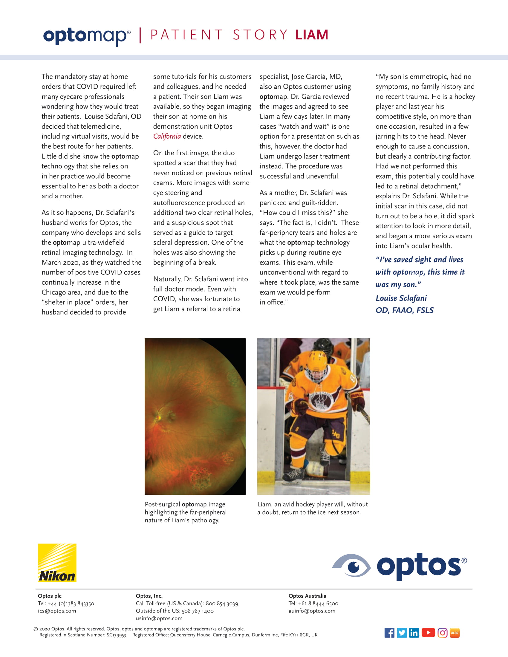
Louise Sclafani, OD, amidst mandatory stay-at-home orders during early 2020, decided that telemedicine, including virtual visits, would be the best route for her patients. Dr. Sclafani’s husband happens to work for Optos, the company that develops and sells the optomap UWF retinal imaging technology, and he decided he wanted to provide some tutorials for his customers and colleagues, finding himself in need of a patient. Their son Liam was available, so they began imaging their son at home on his demonstration unit Optos California device.
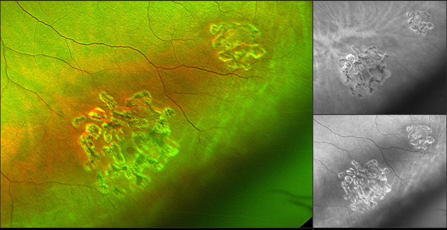
On the first image, the duo spotted a scar that they had never noticed on previous retinal exams. More images with some eye steering and autofluorescence produced an additional two clear retinal holes, and a suspicious spot that served as a guide to target scleral depression. One of the holes was also showing the beginning of a break.
|
Naturally, Dr. Sclafani went into full doctor mode, she was able to quickly obtain a referral to a retina specialist. The doctor had Liam undergo laser treatment rather than a “watch and wait” approach, and the procedure was successful. Liam is an avid hockey player and his competitive nature did however on more than one occasion result in a few jarring hits to the head. Never enough to cause a concussion, but a contributing factor. Had the couple not performed this exam, this potentially could have led to a retinal detachment. While the initial scar in this case, did not turn out to be a hole, it did spark attention to look in more detail, and began a more serious exam into Liam’s ocular health. Read Liam's whole story here. On the first image, the duo spotted a scar that they had never noticed on previous retinal exams. More images with some eye steering and autofluorescence produced an additional two clear retinal holes, and a suspicious spot that served as a guide to target scleral depression. One of the holes was also showing the beginning of a break.
An eye exam is important in any eye trauma or injury to check for changes such as Liam’s and many other traumas that eye care professionals can diagnose and treat before they affect vision permanently. The best solution is always prevention; talk to your eye care professional about including optomap as part of your next eye exam – optomap helps doctors to discover and document the retina with little or no face-to-face interaction and takes only seconds to get a highly-detailed view of the retina, which is critical for detection and management of both ocular and systemic changes.