This month, Optos joins Prevent Blindness America in observing Cataract Awareness Month to further the education surrounding cataracts, what you should know, as well as the value of ultra-widefield (UWFTM) imaging for practitioners as a complement to standard approaches for a comprehensive evaluation of retinal health prior to, and following cataract surgery.
Presently, cataracts are the world’s leading cause of vision loss, accounting for approximately 42 percent of all cases. In the United States, more than 25 million Americans are estimated to have cataracts, according to the report: “Future of Vision: Forecasting the Prevalence and Costs of Vision Problems”. As the population in America continues to age, the number of cataract cases are projected to increase by 50 percent to 38.5 million, by 2032.
What are Cataracts?
Inside our eyes, we have a natural lens. The lens refracts light rays that come into the eye to help us see. The lens should be clear. When cataracts are present, the lens becomes cloudy much like looking through a foggy or dusty car windshield. Things look blurry, hazy or less colorful with a cataract.
Vision changes you may notice if you have a cataract:
- Having blurry vision
- Seeing double
- Extra sensitive to light
- Having trouble seeing well at night, or needing more light while reading
- Seeing bright colors as faded
What Causes Cataracts?
The exact causes of a cataract are unknown, however, most often they are a part of getting older. There are some key possible risk factors that have been identified in those at risk, such as:
- Intense heat or long-term exposure to UV rays from the sun
- Certain diseases, such as diabetes
- Inflammation in the eye
- Hereditary influences
- Long-term steroid use
- Eye injuries
- Eye diseases
- Smoking
There are three different types of cataracts, named according to their locations:
Nuclear cataracts grow in the nucleus (inner core) of the eye’s lens. This is the most common type of cataract associated with aging.
Cortical cataracts develop in the cortex (outer section of the lens).
Posterior subcapsular cataracts form toward the back of a cellophane-like capsule that surrounds the lens. These are most common in people who are diabetic, overweight or taking steroids.
Cataracts can also be classified by cause:
- Age-related cataracts form as result of aging.
- Congenital cataracts occur in babies who are born with cataracts as a result of an infection, injury or poor development before birth. They can also develop during childhood.
- Secondary cataracts are a result of other medical conditions, such as diabetes, or exposure to toxic substances, certain drugs (such as corticosteroids or diuretics), ultraviolet light or radiation.
- Traumatic cataracts develop as the result of an injury to the eye.
Cataracts usually form in both eyes, but not at the same rate. They can develop slowly or quickly, or progress to a certain point, then not get any worse. As a result, one may not notice substantial changes in their sight. Sometimes they can significantly precede symptoms and can be so subtle as to go unnoticed without a comprehensive eye exam.
optomap technology is being increasingly utilized in cataract surgery clinics for immediate views of the retina pre- and post- operatively. optomap images have also proven useful in the process of educating patients about their condition. The UWF view can be captured through problematic, hazy media, where white light has difficulty, revealing any retinal issues that might be a concern prior to surgery, as well as, following surgery. The ability to quickly and easily observe and document retinal health before and after cataract surgery provides both the patient and practitioner a tremendous peace of mind.
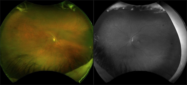
Pre-surgical cataract exam; optomap imaging (color and sensory view) reveals early
Diabetic Retinopathy evident as multiple dot hemorrhages
A published paper from Assil Eye Institute and Batra Vision in California discusses the role of UWF imaging as a standard assessment tool in a cataract procedure. Evaluating the retina prior to, and after, surgery is critical for optimal outcomes. Being able to identify any pathologies before that might adversely affect or delay surgery, as well as, the high expectations for sustained positive visual outcomes from the younger demographic opting for surgery makes a thorough examination increasingly important. The rising number of procedures in already busy clinics further creates a need for a quick and comprehensive evaluation method.
Drs. Kerry Assil MD and Nicholas Batra MD, demonstrate how integrating optomap UWF retinal imaging in a cataract practice can address the demands that come with an increase in procedures. The use of standardized optomap images, within and between practices, reduces variability and facilitates diagnostic consensus. Practice flow is dramatically streamlined because optomap captures more than 80% or 200 degrees of the retina in a single capture in less than ½ second. This process can be administered by a technician, and the digital image is instantly available for review, enabling the doctor to quickly assess and identify if an additional dilated fundus exam is necessary. The optomap image enhances documentation of retinal pathology, cataract opacity, implant choice and postoperative results. Additionally, the efficiency and ease of use of optomap facilitates potential economic advantages through the ability to evaluate a larger population of patients over a shorter period of time.
Integration of Optos UWF retinal imaging into preoperative assessment and postoperative follow-up protocols helps address the issues of an increasing cataract surgery population and heightened patient expectations about postoperative outcomes and extended visual acuity. Optos UWF retinal imaging is an efficient, economical and patient-friendly evaluation tool that provides a consistent point of reference between clinicians and helps to document retinal changes over time and protect the long-term quality of vision.
For more information on Optos UWF retinal imaging, or to hear firsthand the benefits of optomap imaging in cataract practices visit the Optos website.